
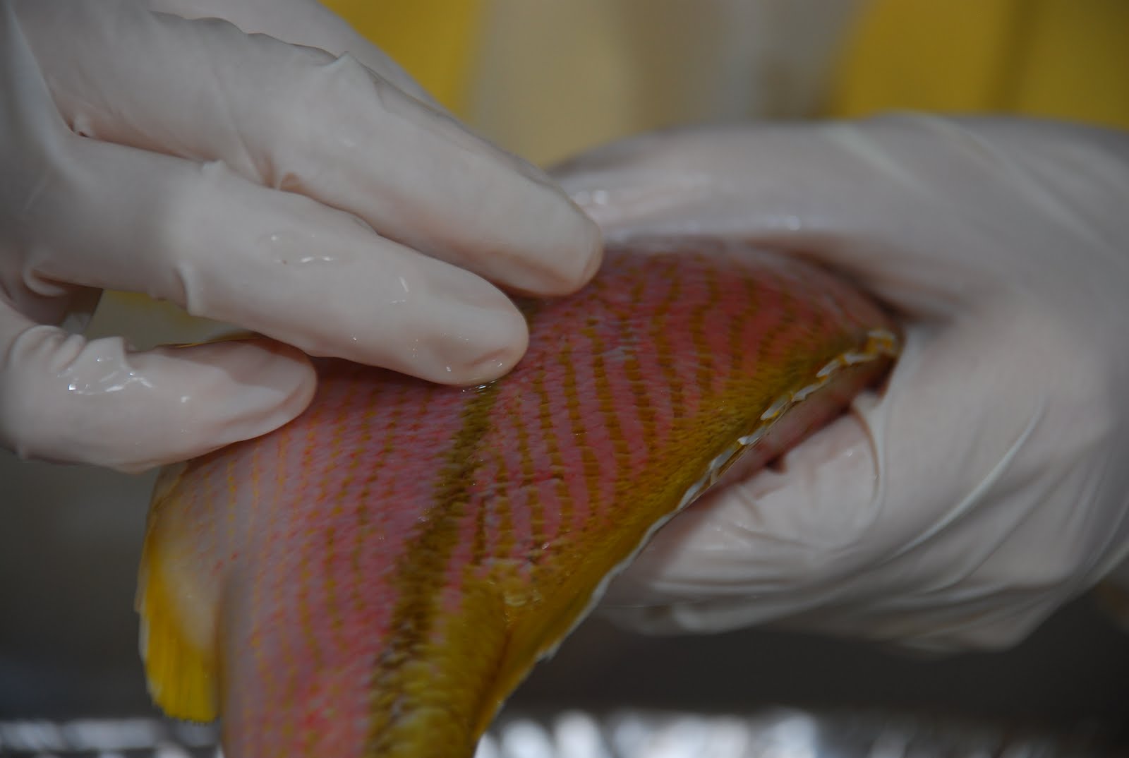
Locate the lateral line.
The lateral line enables the fish to respond to pressure.

Teeth: small, sharp, located in both upper and lower jaws.
Its teeth are small as it does little chewing.
Examine the tongue.
It is non-muscular and is attached to the floor of the mouth.
Fish usually swallow food, therefore a large and elastic esophageal opening is necessary.

Raise the operculum and with a probe carefully separate the layers of gills to examine them. On each side, there are four layers of gills.
The gill chamber is joined to the mouth to enable water to enter the mouth and flow over the gills.
With scissors, cut the left operculum away and remove one set of gills by cutting the upper and lower attachments of the gill arch. Rinse the gills and place them in a culture dish filled with water to be examined later.

Open the body cavity.
Hold the fish with ventral side up, head pointing away from you. Insert your scissors through the body wall anterior to the anus; cut along the midline of the body to the area between the gill covers on the lower side of the head.
Lay your fish on its right side. Continue the incision from the point between the gills, around the front of the operculum, to the top of the body cavity.
Make another incision close to the anus and cut dorsally to the top of the body cavity (see above photo).
With your scalpel, make the remaining incision across the body cavity (just dorsal to the air bladder).
Remove the side wall of the body. The removal of the wall will reveal the body cavity with the organs in their normal positions.

Locate the digestive system.
Find the liver. It is in the anterior end of the body cavity. Gently raise the lobes of the liver and find the gallbladder attached to the lower surface of the liver. Cut the liver free and remove it in one piece.
Locate the short esophagus and stomach.

Locate the air bladder along the top of the body cavity. It may have broken when you removed the body wall.
On the ventral side of the body cavity, near the opercula, locate the pericardial cavity (not pictured), which contains the heart.
The soft upper chamber of the heart is the atrium.
Below and anterior to the atrium is the ventricle.
A purplish muscular bulb, the bulbus arteriosus, gives rise to the ventral aorta, which branches to the gills.

Examine the gills closely.
Examine the feathery filaments and the comblike rakers.
The gill is richly supplied with blood as the exchange of gases in the blood occurs here.
No comments:
Post a Comment